Hair Cross-Section Cortex Observation and Cuticle Laminar-Layer Evaluation
There are a number of objectives when observing hair, including assessing damage from perms or dyeing, or the penetration of hair care products. In particular, the cuticle is most susceptible to such processing, so observing the cross-section of hair in high definition is essential to evaluation. Here, we explain the structure and components of hair, the issues with conventional observation methods, and how they can be solved.
Hair structure
头发是由角蛋白制成的,即角质化蛋白。它对应于角质层或角质层和皮肤上的死皮细胞。轴是头发的可见部分,完全由死细胞组成,这意味着一旦损坏,它们就不会再生。
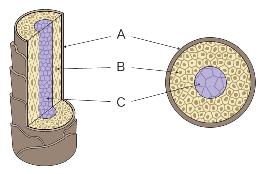
The hair shaft consists of three layers, from outermost to innermost: cuticle, cortex, and medulla.

- A
- Cuticle
- B
- 皮质
- C
- 髓质
Cuticle
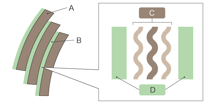
The cuticle is composed of overlapping cells that cover the surface of the hair shaft like scales. One cuticle cell is 0.5 to 1 μm thick, clear and colorless, with moisture-repellent properties. Cuticle cells are aligned in the same direction—you can smoothly run your fingers through your hair from the root, but may feel some scratchy resistance if you try doing this in reverse.
一个角质层细胞覆盖了头发轴周长的约1/3至1/2。如果您的头发僵硬,则可能有更多的角质层细胞覆盖圆周。角质层细胞之间的间隙充满了细胞膜复合物(CMC)。
CMC由将三角层(蛋白质)夹的β层(脂质)组成。厚度为0.04至0.06μm。染发剂和其他发型产品通过CMC穿透头发。

- A
- Cuticle
- B
- CMC
- C
- 三角洲层(蛋白质)
- D
- β层(CMC脂质)
皮质
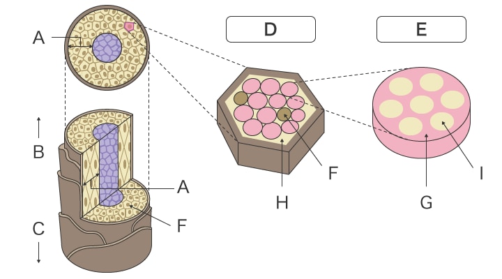
皮质是一组宏观纤维。每个宏观纤维都是微纤维的组件,它们之间的间隙充满了具有补充特性的亲水基质。该基质的主要成分是无定形角蛋白。它含有丰富的黑色素色素,以及氨基酸,多肽(PPT),核酸和矿物质。矩阵是受珀姆斯影响或由染发剂染色的部分,因此对护发很重要。

- A
- 皮质
- B
- 头发的尖端
- C
- Root of hair
- D
- 皮质细胞
- E
- Macrofibril
- F
- Melanin
- G
- Matrix
- H
- Amorphous keratin
- I
- Microfibril
髓质
髓质位于发轴的中心,由由蛋白质和脂质制成的细长立方细胞组成。它在细胞之间包含空气,并且该缝隙反映了光。当这个差距扩大时,头发显得变白了。
并非所有的头发都有髓质。薄的头发和细发没有髓质。在某些地方也可能是不连续的。
头发的高分辨率观察
Optical microscopes or scanning electron microscopes (SEM) are used for high-resolution observation of hair.
显微观察是镜面ref下完成的lection light by observing hair from the side using coaxial illumination. Observation of flat surfaces under specular reflection light is bright due to the reflected light directly entering the lens or CCD. In contrast, slopes in stepped parts appear darker due to some reflected light escaping off the surface. This contrast allows for fine structural visualization of the surface of hair. However, the surface of hair is curved, and reflected light escapes outward even more on the edges of the hair. Cross-section evaluation using an optical microscope does not provide the resolution necessary to observe the laminar structure of the cuticle, which may result in images of a single cuticle layer.
On the other hand, while a scanning electron microscope (SEM) allows for high-resolution observation of the cuticle surface structure, observation of non-conductive specimens requires pretreatment to prevent charge-ups, in which the incident electron beam accumulates on the surface of the specimen and creates a foggy glow. This can take a lot of time and effort. In addition, the hair must be placed inside a vacuum chamber for observation under vacuum, which causes the moisture in the hair to evaporate and may change the hair’s morphology. Whichever approach is used, it may be difficult to observe hair in its natural state.
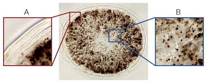
With the high-resolution offered by our BZ-X800 All-in-One Fluorescence Microscope, you can clearly observe specimens with laminar structures like the cuticle.You can also observe the melanin spread across the cortex in clear view, and simultaneously evaluate the cortex and cuticle.

- A
- Laminar structure of cuticle
- B
- 黑色素在皮质中发现
- 样本
- Cross-section of human hair
- 成像放大率
- 物镜镜头100倍(浸入油)
- 成像方法
- 布莱特菲尔德
- Using the All-in-One Fluorescence Microscope BZ-X800
-
- 使用BZ-X800可以在正常观察模式下进行完整的角质层观察和荧光显微镜。
- 通过高分辨率,横截面观察,您可以同时评估皮质中表皮和黑色素的层状结构。
- Combined with fluorescent labeling, you can also observe the penetration of hair care products into the hair. The BZ-X800’s ability to quantitatively analyze multiple samples supports quantified evaluation.
- Here are some examples of using the All-in-One Fluorescence Microscope BZ-X800 in front-line research.
- [Myelodysplastic Syndromes (MDS)] Stitching, Sectioning and the Z-Stack Function as Decisive Arguments for the Acquisition of the BZ Fluorescence Microscope at the University Hospital of Düsseldorf
- [Neuropathology] The perfect solution for everyday patient diagnostics and clinical research at the Institute of Neuropathology in the Charité hospital in Berlin
- [再生医学] BZ系列为神经干细胞和脊柱观察提供了必需的成像
- [Gene Therapy] Improving Research for the Development of Gene Therapy Drugs
- [Heart Disease Treatment] Developing Cell Sheets for Myocardial Regenerative Treatments
- [Cancer Treatment] Automated Fluorescence Microscope Transforms Process for Induced Cancer Stem Cell Research
- [Immune System] BZ Series Contributes to Understanding the Pathological Model of Asthma
- [Biomaterials] Promoting Efficiency in Research With Compact, User-friendly Microscopes


